| Description: |
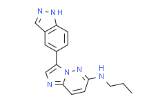
CHR-6494 is a potent inhibitor of haspin, inhibiting histone H3T3 phosphorylation, with an IC50 of 2 nM. |
| Target: |
IC50: 2 nM (haspin)[1] |
| In Vivo: |
CHR-6494 (50 mg/kg, i.p.) inhibits the growth of tumor and cuases no obvious body weight change in nude mice bearing HCT-116 human colorectal cancer cells[1]. |
| In Vitro: |
CHR-6494 is a potent inhibitor of haspin, inhibiting histone H3T3 phosphorylation, with an IC50 of 2 nM. CHR-6494 does not modify H3S10 and H328 phosphorylation levels, and shows no significantly inhibitory effects on other protein kinases such as Aurora B kinase. CHR-6494 dose-dependently inhibits the growth of cancer cells, such as HCT-116, HeLa, MDA-MB-231, and Wi-38 cell, with IC50s of 500 nM, 473 nM, 752 nM and 1059 nM, respectively. CHR-6494 (500 nM) produces a mitotic catastrophe with abnormal morphology of the mitotic spindle and centrosome amplification, and upregulates the spindle assembly checkpoint protein BUB1 and the marker of mitotic arrest cyclin B1[1]. CHR-6494 exhibits inhibitory activities against melanoma cell lines, including BRAFV600E mutants, NRAS mutants, and wild type cells, with IC50s ranging from 396 nM to 1229 nM. CHR-6494 (300 nM and 600 nM) induces apoptosis, increases caspase 3/7 activity by 3- and 6-fold, respectively in COLO-792 cells, and to 8.5- and 16-fold in RPMI-7951 cells. CHR-6494 in combination with MEK inhibitors synergistically inhibits viability of melanoma cells, enhances apoptosis in melanoma cells, modulates cell cycle progression independently by arresting melanoma cells at different phases, and suppresses migration of melanoma cells[2]. |
| Kinase Assay: |
The analysis of the enzymatic inhibitory capacity of the compound in a panel of 29 protein kinases is developed using a FRET assay based on the differential sensitivity of phosphorylated and non-phosphorylated peptides to protein cleavage (Z′-LYTE Kinase Assay). In the primary reaction, the kinase transfers the γ-phosphate of ATP to a single tyrosine, serine or threonine residue in a synthetic FRET peptide. In the secondary reaction, a site-specific protease recognizes and cleaves non-phosphorylated FRET peptides. Phosphorylation of FRET peptides suppresses cleavage by the development reagent. Cleavage disrupts FRET between the donor (coumarin) and the acceptor (fluorescein) fluorophores on the FRET peptide, whereas uncleaved, phosphorylated FRET peptides maintain FRET. A ratiometric method, which calculates the ratio (the emission ratio) of donor emission to acceptor emission after excitation of the donor fluorophore at 400 n, is used to quantitate reaction progress[1]. |
| Cell Assay: |
Cells are treated for 24, 48 and 72 h with the inhibitor or with DMSO as a control. Cell viability is assessed using the colorimetric XTT assay. Cells are seeded in 94-well plates at a density of 4 × 10⁴ cells per well, and allowed to attach for 24 h. The medium is then exchanged with others containing different drug concentrations (0.001−10 μM). Eight wells for each concentration of the CHR-6494 compound are used. At the corresponding time, the culture medium is discharged, the XTT reagent is added and the final cell number and optical density are determined. Dose-response curves are generated and cell viability is evaluated after 72 h of treatment. The half-maximal inhibitory concentration (IC50) is determined using GraphPad Prism software[1]. |
| Animal Administration: |
Athymic nu/nu male mice, aged 4-5 weeks, are used for tumor xenograft assays. Animals are maintained in a sterile environment; their cages, food and bedding are sterilized by autoclaving. Mice are anesthetized and tumor cells are injected subcutaneously. In all, 3.5 × 10⁶ exponentially growing HCT-116 cells diluted in 250 μL of sterile PBS are injected subcutaneously in each animal (n = 30). Body weight is recorded and tumor dimensions are measured twice weekly using digital calipers. Tumor volume (in mm3) is estimated according to the formula V = D × d2/2, where D is the long axis and d the short axis of tumor. When tumors reach an average volume of 200 mm3 (15 days after injection), 24 mice harboring homogeneous tumor sizes are randomized into two groups: (1) control group (n = 8) treated with vehicle (solution of 10% DMSO/20% 2-hydroxypropyl-b-cyclodextrin; (2) treatment group (n = 16) mice is diary treated by intraperitoneal injection of 50 mg/kg of CHR-6494 diluted in a solution of 10% DMSO/20% 2-hydroxypropyl-b-cyclodextrin in two cycles of five consecutive days for 15 days. The treatment group is randomly divided into a short-time response group (n = 8), defined by tumor weight at the moment of killing of the control group, and a long-time response group (n = 8), defined by tumor regrowth after treatment. Mice are killed at the end of treatment, and tumors from both groups are excised and weighted. The mean volume of tumor mass is expressed as mean ± s.e.m. for each mouse group, and significance is assessed by means of the Mann-Whitney U-test. Values of P < 0.05 are considered statistically significant. Upon killing mice, colon, lung, liver and kidney tissues are obtained to analyze endogenous toxicity by hematoxylin and eosin[1]. |
| References: |
[1]. Huertas D, et al. Antitumor activity of a small-molecule inhibitor of the histone kinase Haspin. Oncogene. 2012 Mar 15;31(11):1408-18.
[2]. Han L, et al. Anti-Melanoma Activities of Haspin Inhibitor CHR-6494 Deployed as a Single Agent or in a Synergistic Combination with MEK Inhibitor. J Cancer. 2017 Aug 25;8(15):2933-2943. |
 To enhance service speed and avoid tariff delays, we've opened a US warehouse. All US orders ship directly from our US facility.